About the cross-section plates
If you have a basic or full knee kit model, your kit comes with cross-section plates attached to either end of the kit.
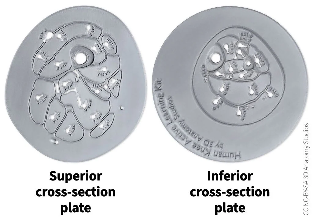
The cross-section plates included with the basic and full knee kit models.
The plates show you a cross-section of the anatomy (in the transverse plane) at the middle of the thigh (superior) and middle of the leg (inferior), including the muscles, arteries, veins, nerves, and skin line. The arteries, veins, and nerves are represented by holes in the plate. The shapes and sizes of all the structures in the cross-section plates are accurate anatomical representations, coming directly from the same cryosection data that was used to make the kit. Having these plates "bookend" the knee kit relates the 3D structures of the knee kit with a 2D cross-sectional representation (as seen in a CT slice, for example) and connects the knee kit with the structures just outside of the knee region.
The image below identifies the structures in the superior cross-section plate.
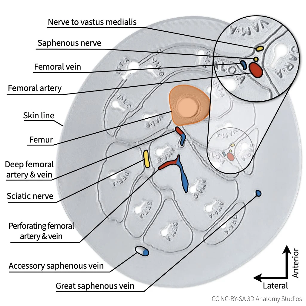
The superior cross-section plate with labels. Muscles are not labeled since they are already identified by the abbreviations printed into the plate.
And here is the superior cross-section plate with the corresponding slice in the cryosection data.
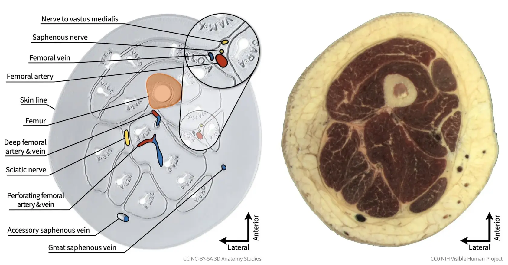
The superior cross-section plate (left) side by side with the corresponding cryosection (right). Note that the entire edge of the superior cross-section plate represents the skin line, with the space between the skin and muscles being filled primarily with subcutaneous adipose tissue.
The image below identifies the structures in the inferior cross-section plate. For the superior cross-section plate, the entire outer edge represents the skin line. However, for the inferior cross-section plate, the plate is much larger than the leg itself (expanded outward to provide a larger base for stabilizing the knee kit) and the skin line is represented by a single, continuous indented line.
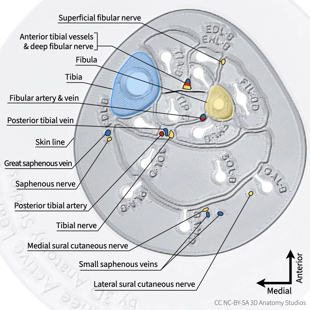
The inferior cross-section plate with labels. Muscles are not labeled since they are already identified by the abbreviations printed into the plate. The area of the plate outside of the skin line has been grayed out and the image zoomed in.
And here is the inferior cross-section plate with the corresponding slice in the cryosection data.
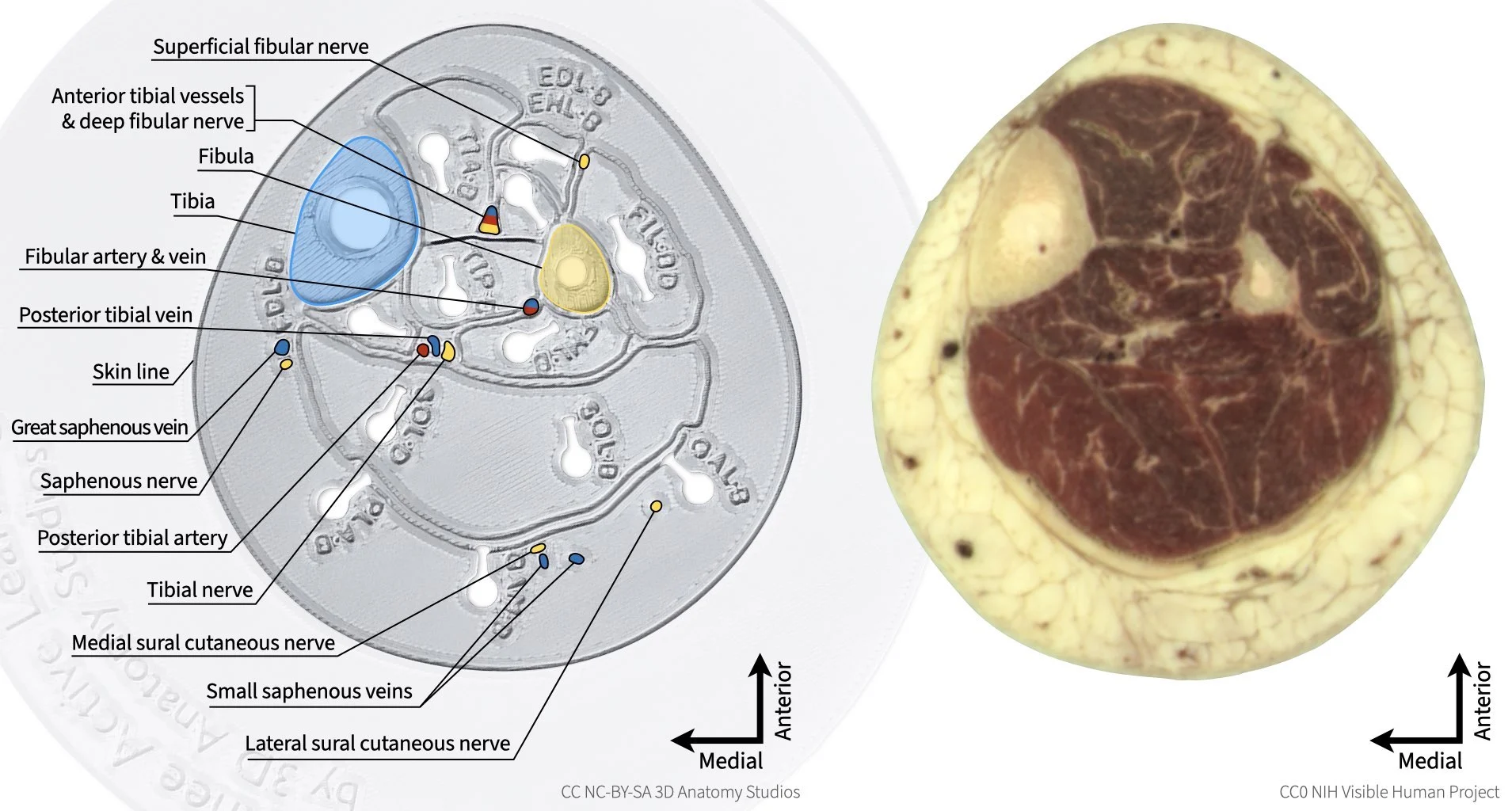
The inferior cross-section plate (left) side by side with the corresponding cryosection (right).